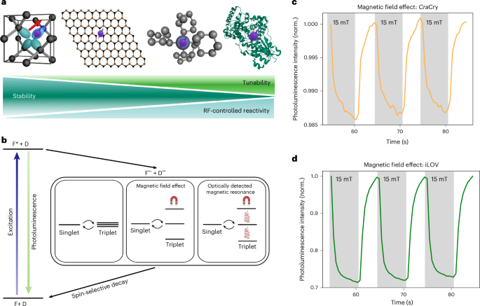
Solid-state spins in semiconductors, such as the nitrogen-vacancy center in diamond1 or boron vacancy in hexagonal boron nitride2, are the workhorses of quantum sensing3 (Fig. 1a). Their spin–photon interface enables optically detected magnetic resonance (ODMR) for various sensing applications in biology and physics. Despite their useful properties, synthetic tunability of their optical and spin properties or deterministic fabrication methods have remained elusive4. Molecular spin systems offer a compelling alternative, allowing bottom-up design with two key advantages: tunability through precise atomistic control over the molecular structure and its associated properties and scalability through chemical assembly. Recently, the synthesis of organometallic molecules with an optical spin interface has been demonstrated5. Here, we show that certain flavoproteins also exhibit optically addressable spin states. The main advantage of the genetically encoded protein scaffolds is their biocompatibility and their tunability using rational design6 or directed evolution7.

a, Overview of various spin systems, including the nitrogen-vacancy center in diamond, the boron vacancy in hexagonal boron nitride, organometallic molecules and, as demonstrated in this work, proteins. The degree of tunability increases from semiconductor-based systems to biological systems. Radical pairs and their yields in chemical and biological systems can be controlled by RF fields. b, Simplified schematic of the photophysical processes and spin dynamics of a photoinduced SCRP based on flavin (F) and a donor (D). The interconversion between triplet and singlet states can be influenced by magnetic fields (MFE) and/or RF fields (ODMR), thereby modulating the decay through spin-state-selective channels. c, MFE (~1.5%) observed in CraCry. d, MFE (~30%) observed in iLOV.
Photoactive flavin-containing proteins, such as cryptochromes (Crys) and light–oxygen–voltage (LOV) domain proteins, respond to blue light by generating spin-correlated radical pairs (SCRPs) involving the flavin cofactor and a nearby amino acid residue (Fig. 1b). These radical pairs are initially formed in a distinct spin-correlated singlet or triplet state8. Spin interconversion between these states, governed by hyperfine interactions and influenced by external magnetic fields, can modulate the recombination kinetics and ultimately affect the photochemical outcomes of the reaction9. In LOV proteins, these SCRPs only form if the natural reaction is blocked by a mutation, in which case they are not involved in signaling10,11. Crys, however, are believed to have a direct role in magnetoreception, for example, enabling birds to navigate using the Earth’s magnetic field8,12,13.
Here, we demonstrate that these proteins exhibit ODMR under ambient conditions. In parallel to our work, ODMR in engineered LOV proteins (MagLOV)14, in red fluorescent protein–flavin systems15 and in fluorescent protein systems16, the latter of which does not rely on SCRPs, has been shown. Our complementary work shows that radical-pair-based ODMR also occurs in the archetypal Crys17 and in improved LOV (iLOV)18, a well-established fluorescent reporter.
In the first step, we measure the photoluminescence (PL) of the animal-like Cry from the green alga Chlamydomonas reinhardtii (CraCry) as a function of the applied magnetic field strength B0 (Earth’s magnetic field and 15 mT) (Fig. 1c). We observe that the PL intensity is affected by the presence of a magnetic field—a phenomenon known as magnetic field effect (MFE). This effect can be attributed to the modulation of the singlet–triplet interconversion of the formed SCRP12,19,20,21 (Fig. 1b), which alters the concentration of the different flavin species (mainly the oxidized flavin) under the given experimental equilibrium conditions. We also include a LOV protein (specifically iLOV) in our studies, which is a domain that has been optimized to function as a fluorescent marker in cellular imaging18,22. Similarly, we observe an MFE, albeit with greater contrast (Fig. 1d), which is laser excitation power dependent (Extended Data Fig. 1). The negative MFE (that is, the reduction in PL with applied magnetic field) indicates a triplet-born radical pair in both cases23. While this is consistent with LOV proteins, the results differ from established data on Cry, which indicate a singlet-born radical pair24,25. This suggests that, under our experimental conditions, alternative radical pairs may be formed26. Details and reference experiments can be found in Supplementary Note 1.
While MFEs have been studied in detail previously12,20,27, it inspired us to perform ODMR experiments, similar to their optically active solid-state spin counterparts28. In these experiments, radio wave or microwave frequencies (RF) are used to address spin transitions that couple to the optical transitions which can be read out from the PL intensity. Therefore, we developed a modified ODMR setup29 that includes temperature-controlled sample handling, a tunable magnetic field B0 and electronics for RF delivery, combined with sensitive PL detection (Methods and Fig. 2a). A typical ODMR pulse sequence is shown in Fig. 2b, where we apply a specific RF and simultaneously record the PL intensity. To cancel noise (for example, laser intensity fluctuations), a reference measurement is recorded without the RF pulse. Typically, in ODMR experiments, the magnetic field is held constant while the RF is swept29. We avoid this approach here, as it can lead to unwanted modulation of the resonance lineshape because of the frequency-dependent characteristics of the RF delivery structures (Supplementary Note 2). Instead, we keep the RF fixed and sweep the magnetic field strength. We use a previously characterized solid-state spin system (S = 1/2) in boron nitride nanotubes (BNNTs)30,31 for calibrating the magnetic field strength in our experiment (Supplementary Note 3).

a, Experimental setup of the ODMR experiments. b, Continuous ODMR pulse sequence. The pauses between successive experiments provide time for sample recovery. c, ODMR spectrum of CraCry recorded at an excitation frequency of 1,470 MHz. Inset, CraCry crystal structure (PDB 5ZM0) including the FAD cofactor and the electron-transfer chain (orange). d, CraCry PL spectra under RF irradiation. e, ODMR spectra of CraCry recorded under different RF excitation frequencies. f, The dependence of the CraCry resonance frequency on magnetic field strength is consistent with a g ≈ 2 spin system. g, ODMR spectrum of iLOV recorded at a RF excitation frequency of 1,470 MHz. Inset, iLOV crystal structure (PDB 4EET) including the FMN cofactor and aromatic amino acids (orange). h, iLOV PL spectra under RF irradiation. i, The ODMR contrast of iLOV as a function of the readout time indicates a slow buildup, reflecting slow underlying chemical kinetics. The objective schematic in a was created in BioRender; Meng, K. https://BioRender.com/qyxrdke (2026).
First, we apply an RF of 1,470 MHz, sweep the magnetic field from 42.5 mT to 62 mT and monitor the PL intensity of the CraCry sample. An enhanced PL intensity is observed around 52.5 mT, corresponding to the expected spin transition of a free electron (Fig. 2c,d). The linewidth of approximately 100 MHz is in good agreement with the linewidth expected from hyperfine interactions32. To verify that the signal indeed originates from an electron spin resonance, we excite the CraCry sample at different RF frequencies and sweep the magnetic field strength in each case (Fig. 2e). The ODMR resonance shifts consistently according to a g ≈ 2 system, confirming that we are observing an electronic spin resonance (Fig. 2f).
Next, we performed ODMR experiments on iLOV. The observed ODMR signals are qualitatively similar to those of CraCry but with an impressive contrast of nearly 50% after optimization (Fig. 2g and Supplementary Note 4). In this case, we also confirm a g ≈ 2 system (Extended Data Fig. 2). We also note the drastically enhanced brightness and stability of iLOV compared to CraCry (Supplementary Note 5). We observe a slightly narrower linewidth (~70 MHz) for iLOV compared to CraCry (~100 MHz), which is independent of 15N labeling (Extended Data Fig. 3 and Supplementary Note 6). PL spectra indicate an increase in the oxidized flavin mononucleotide (FMNox) state upon RF excitation (Fig. 2h). Importantly, the signal builds up over time (Fig. 2i), indicating that, similar to the MFE (Fig. 1c,d), the ODMR contrast arises from a slow shift in the chemical equilibrium of the flavin states driven by spin chemistry9,12. The dynamics are governed by optical excitation and RF power, as well as other factors influencing the chemical equilibrium. The observation is best explained by the formation of long-lived states, for example, generated through redox or (de)protonation reactions, which subsequently decay on the timescale of milliseconds to seconds. Although these states may be only weakly populated after optical excitation, their accumulation under continuous optical and RF excitation conditions leads to a substantial ODMR contrast. In addition, we perform pulsed ODMR experiments in which optical excitation and RF manipulation are temporally separated (Extended Data Fig. 4 and Supplementary Note 7). These measurements demonstrate that RF manipulation is possible during the lifetime of the flavin triplet state observed in transient absorption experiments, which is also the timescale on which SCRPs are being formed. All of these observations point toward the SCRP mechanism8 as a tentative explanation for the observed ODMR effect (Fig. 1b). In the MFE experiments, the applied magnetic field splits the triplet states, which alters the recombination kinetics of the radical pair, ultimately affecting the equilibrium concentration of flavin molecules in the ground state under continuous optical excitation. The applied RF fields induce spin transitions between the T0 and T+/T− triplet states and, thus, reverse the MFE (Fig. 1c,d), leading to an increased PL intensity.
We note that ODMR can also originate directly from triplet states16,33. However, such a mechanism typically does not generate long-lived states on the millisecond-to-second timescale and, more importantly, would be expected to produce a pronounced zero-field splitting in the GHz range34, which is not observed in our data.
The ability to detect ODMR in proteins opens up a wide range of potential applications. Similar to optically active defects in solid-state systems, spin resonance can be used for magnetic field sensing28. Because of its superior ODMR performance, brightness, robustness, compact size and suitability for biotechnological applications, iLOV was selected for subsequent experiments. To demonstrate this, we position a small magnet adjacent to the sample, generating a magnetic field gradient across the microscope’s field of view (Fig. 3a). Unlike in our previous ODMR experiments, we retain spatial information by analyzing the spectrum recorded on each camera pixel. The resulting ODMR resonance shifts across the field of view reflect the local magnetic field variations. In these experiments, we record the ODMR as a function of RF. Although the detailed lineshape and structure of the ODMR signal are affected by the RF delivery, we empirically found that weighting the ODMR contrast allows the extraction of an average resonance frequency, which serves as a reliable calibration metric for the magnetic field (Supplementary Note 8). Applying this fit pixelwise allows us to construct a magnetic field map, visualizing the gradient across the sample (Fig. 3b). We validate the magnetic field gradient with the BNNT solid-state spin system (Supplementary Note 9). This technique offers a promising route for genetically encodable spin-chemistry-based magnetic field sensing, providing a complementary approach to quantum diamond microscopy, which is often limited by the distance between the sensor and the biological sample35.

a, Schematic of the experimental setup for magnetic field sensing. A small magnet placed next to the iLOV sample generates a magnetic field gradient across the microscope’s field of view. b, By fitting the ODMR spectra pixelwise, a spatial map of magnetic field strength is obtained, visualizing the gradient across the sample. c, The magnetic field gradient, combined with RF fields, enables spatially selective excitation of regions that match the spin resonance condition. Depending on the position, the protein has a different ODMR resonance, encoded in the magnetic field gradient shown in d. e, PL detection as a function of RF demonstrates spatial control over PL intensity and, consequently, over spin-dependent radical recombination processes. Each applied RF fRF selectively excites iLOV spin transitions (c) at specific locations that match the ODMR resonance condition, resulting in an increase in PL. The image shows the relative PL change.
Lastly, the combination of RF control and magnetic field gradients enables spatial control of the SCRP in the proteins. Analogous to techniques used in magnetic resonance imaging, a magnetic field gradient B(x) allows us to encode the position into a resonance frequency according to the relation f = γB(x), where γ is the gyromagnetic ratio of the electron (Fig. 3c,d). By applying a specific RF fRF, only the region or ‘slice’ of SCRPs that satisfies this resonance condition is selectively excited. This localized excitation by RF pulses alters the radical recombination yield and, consequently, the PL at that position. Spatial control of PL is visualized by calculating the PL ratio between measurements with RF applied (RF on) and without RF (RF off), which is shown for different frequencies fRF in Fig. 3e. By tuning the RF, we can selectively address proteins in certain regions, where the spatial resolution is determined by the strength of the applied magnetic field gradient.
The ability to modulate the PL intensity of proteins can enhance sensitivity (for example, by suppressing background PL36) or enable super-resolution techniques in which spatial resolution is encoded in magnetic field gradients, thereby surpassing the limits of optical resolution37. Moreover, our results demonstrate that (photo)chemistry of flavin states can be controlled using RF fields—a key distinction from solid-state spin defects, where the ODMR response is governed by photophysical processes (Fig. 1a). This paves the way toward future RF-based control of biological processes such as gene expression or signaling38,39,40,41,42.
In summary, we show that spin states in CraCry and iLOV proteins can be manipulated with RF fields and read out optically at ambient temperature. The inherent tunability of proteins through rational design6 or directed evolution7 offers exciting opportunities for engineering spin–optical interfaces. Because of the high optical contrast, brightness and stability, we anticipate that iLOV in particular will find broad applications across quantum technologies, bioimaging and RF-controlled biochemistry38,39,40,41.